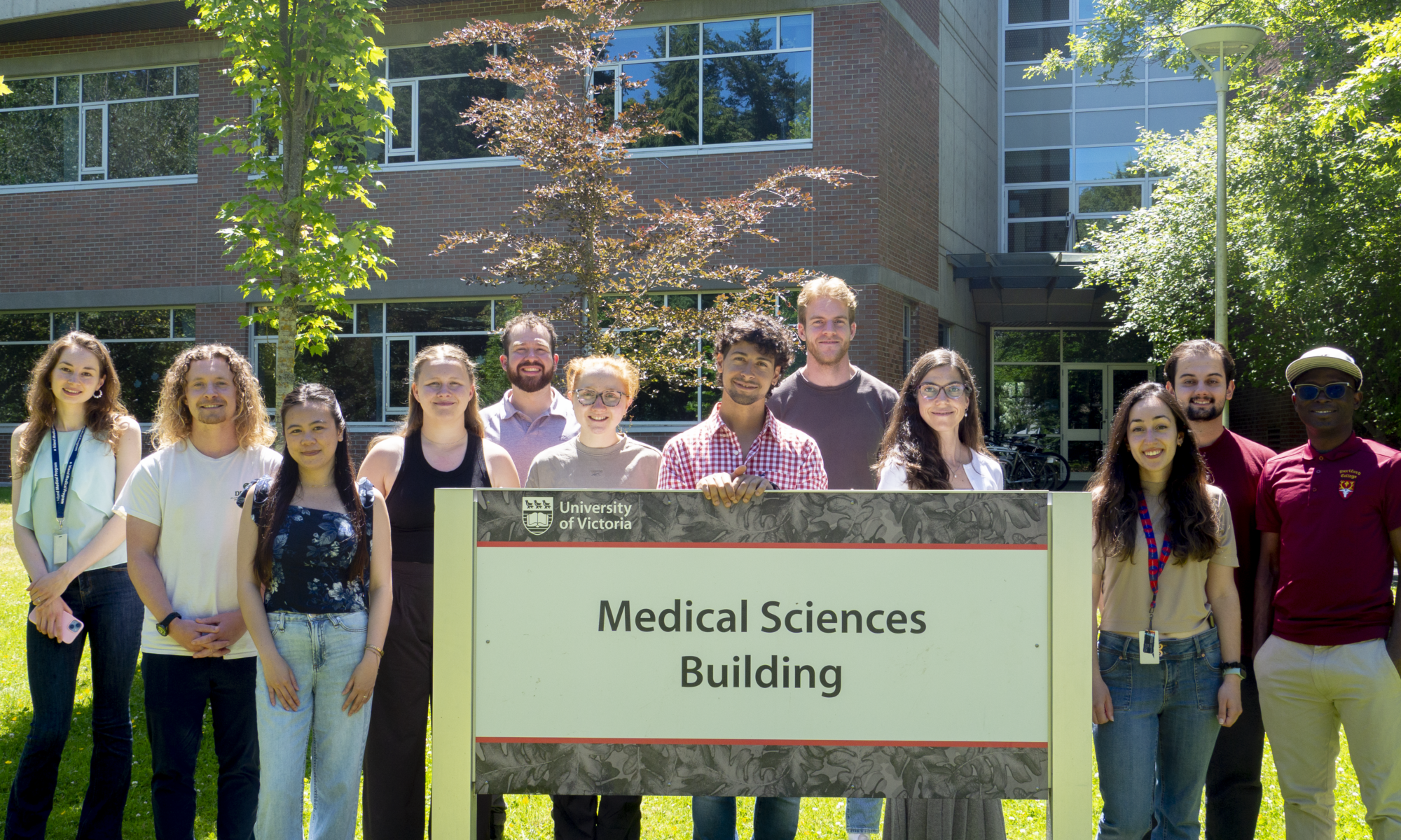

Electron Microscopy Services
Dr. Tremblay has 23 years of experience with biological electron microscopy (EM), from sample preparation to imaging and ultrastructural analysis. With recent developments in the EM field, the Tremblay Lab has transitioned from using transmission EM to scanning EM, significantly expanding the possibilities for both 2D and 3D imaging.
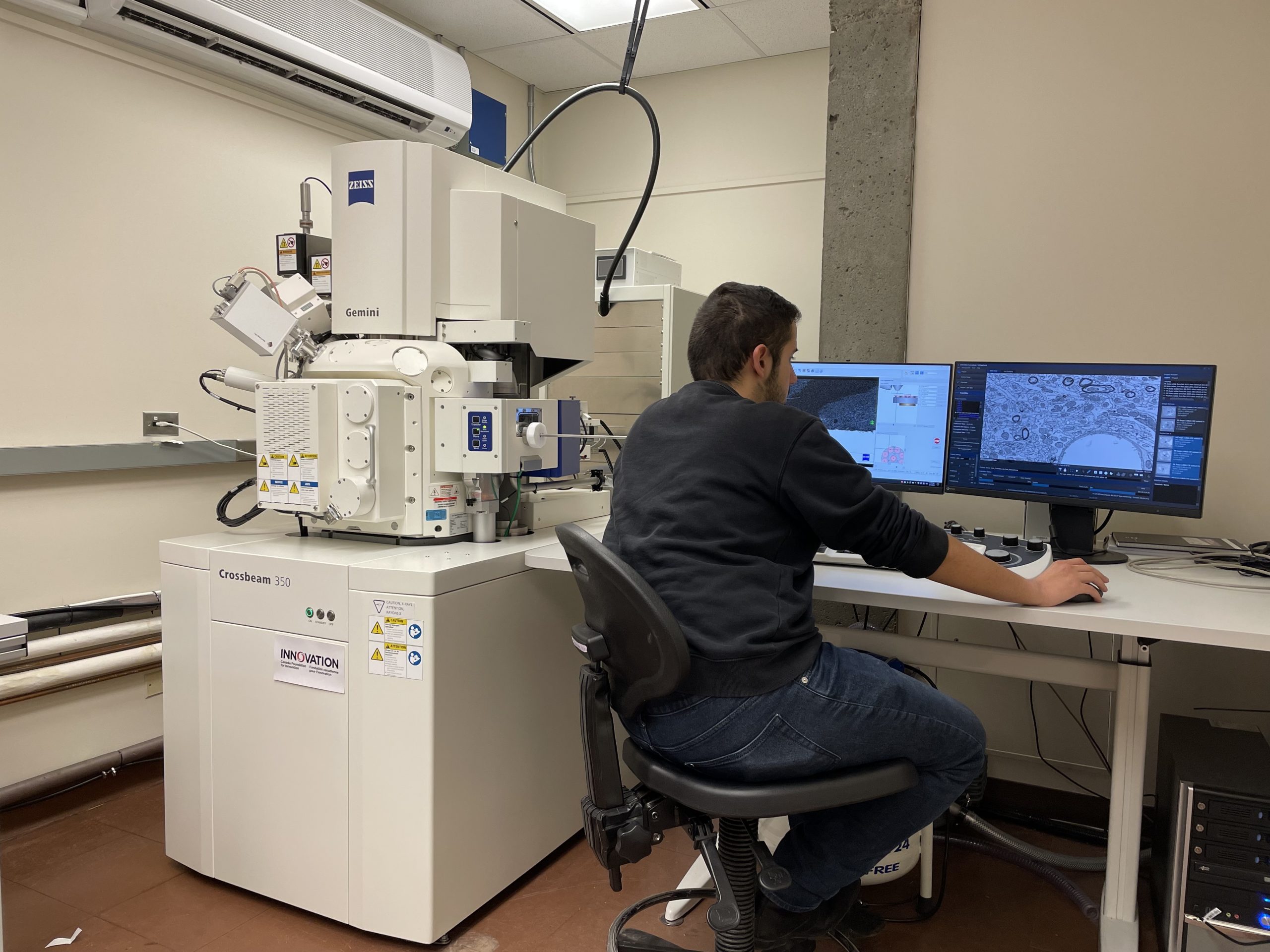
At the University of Victoria, the Tremblay Lab has access to a state-of-the-art Zeiss Crossbeam 350 focused-ion beam (FIB)-SEM system. The Tremblay Lab has developed over the years a unique workflow for semi-automated imaging, both in 2D (chip mapping) and 3D (FIB-SEM), combined with immunostaining. Recently, we have also developed conjugate fluorescence array tomography to expand the number of markers used for cell type and subtype characterization.
This platform has been applied to a wide range of biological samples, including cells, 2D and 3D cellular models, animal models, and human post-mortem samples. While our primary focus and expertise lie in studying brain resilience and cognitive aging, notably with regard to microglia-neuron interactions, we have optimized protocols for oocytes, snails, worms, and various other sample types.
Our extensive collaborations on the national and international levels provide an exchange of expertise and training in advanced EM approaches.
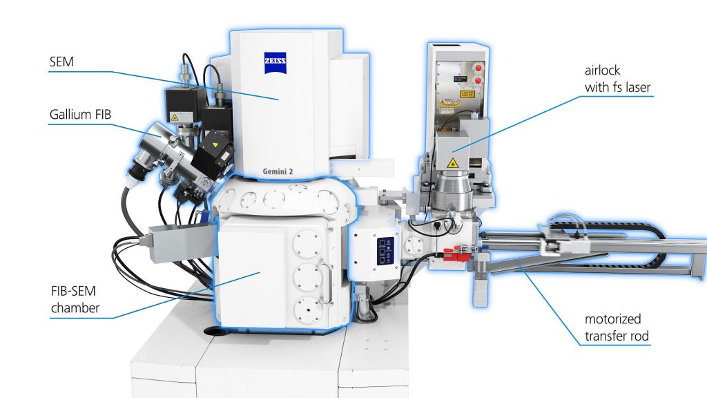
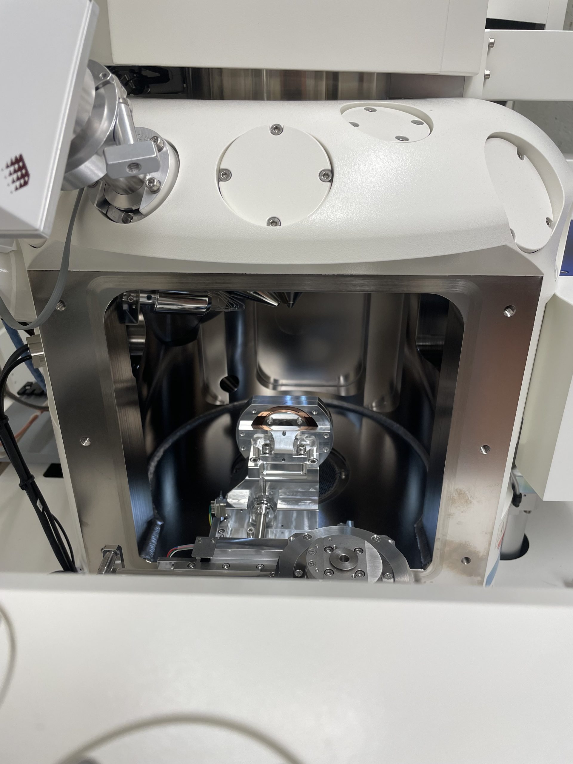
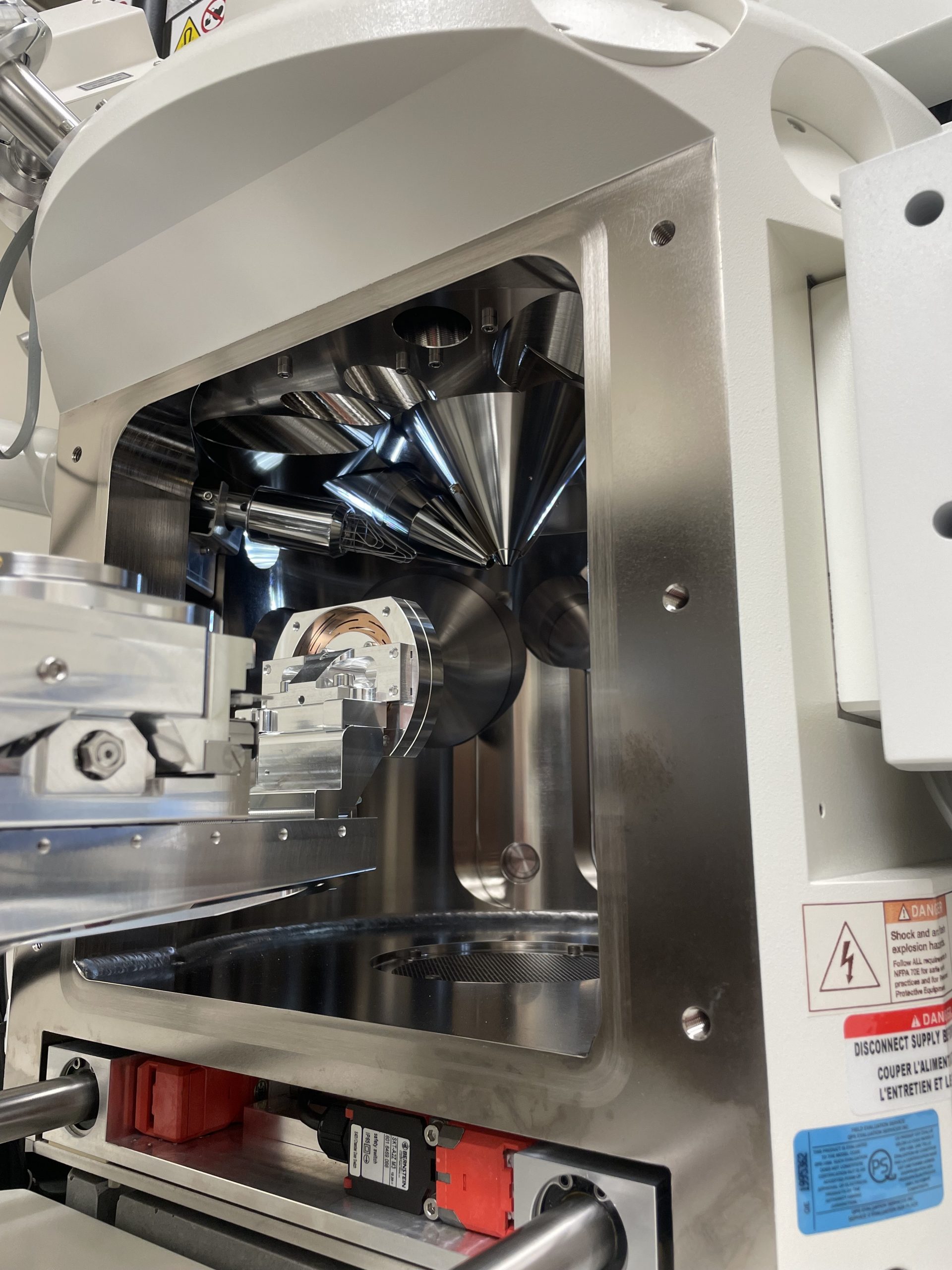
The Zeiss Crossbeam 350 is a state-of-the-art focused ion beam scanning electron microscope (FIB-SEM) that integrates a high-resolution SEM column with a gallium ion beam. This dual-beam configuration allows for precise milling of resin-embedded biological samples while simultaneously imaging the exposed surface. By repeating this layer-by-layer process, the system generates serial images that can be reconstructed into high-resolution three-dimensional datasets. This makes the Crossbeam 350 particularly powerful for ultrastructural studies of cells, organelles, and neural circuits.
Compared to traditional transmission electron microscopy (TEM), the Crossbeam 350 offers significant advantages for volumetric imaging. While TEM provides excellent resolution, it requires manual ultrathin sectioning and alignment. In contrast, the FIB-SEM enables reduced distortion in sectioning with isotropic resolution, allowing a more accurate of the cellular architecture.
The Tremblay Lab’s Zeiss Crossbeam 350 scanning electron microscope was acquired with funding from a Canada Foundation for Innovation John R. Evans Leaders Fund grant (#39965 Laboratory of ultrastructural insights into the neurobiology of aging and cognition).
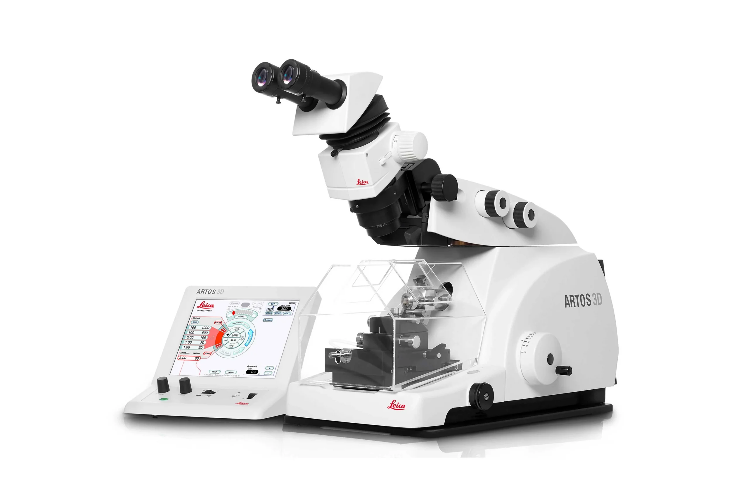
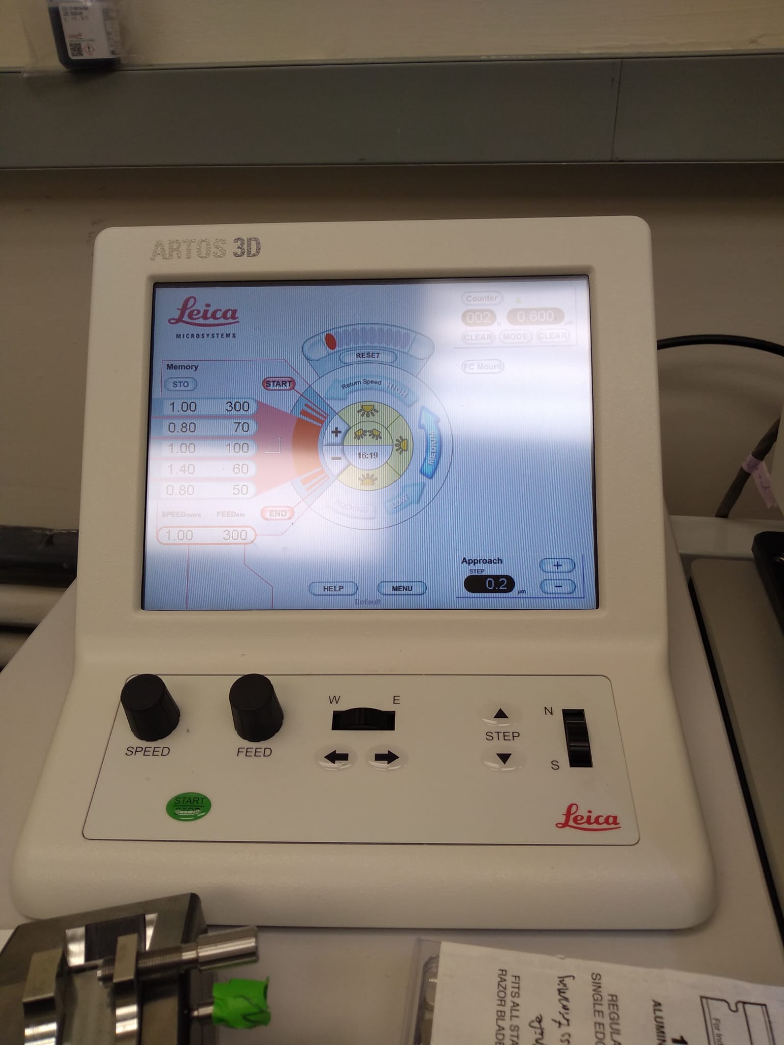
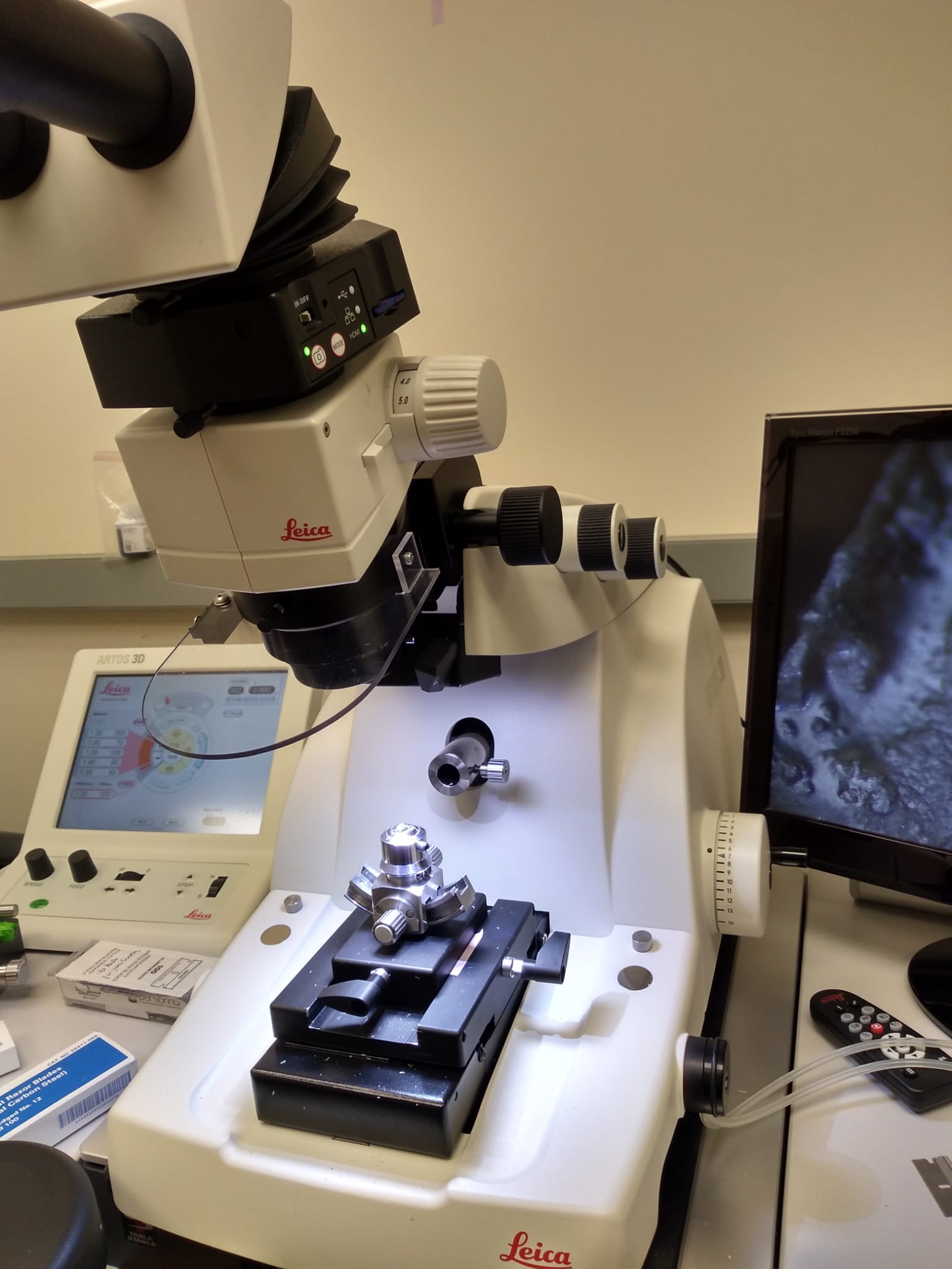
The ARTOS 3D ultramicrotome (Leica Microsystems) is an automated system that produces long ribbons of serial ultrathin sections with high consistency and minimal artifacts, enabling efficient large-volume imaging.
It provides a high-throughput method for volumetric reconstruction: ARTOS generates physical serial sections that are imaged by array tomography on the Crossbeam 350, while FIB-SEM on the same Crossbeam 350 system performs in situ milling and imaging for isotropic datasets. In a correlative workflow, resin-embedded samples are sectioned with ARTOS, mapped to identify regions of interest, and then imaged by array tomography or FIB-SEM for 3D analysis.
How to start a collaboration with us?
- Contact Dr. Tremblay (evetremblay@uvic.ca) with your project summary and number of animals requested for processing.
- Dr. Tremblay will analyze the inquiry and assess priority by putting a timeline together with you.
- We will prepare a quote based on the service requested, which needs to be signed by both parties
- The University of Victoria will prepare an invoice. We usually request a payment of 50% beforehand.
- We will take care of the sample preparation, processing and imaging, according to what has been discussed.
- Results generation and final 50% payment from collaborator.
2013, Université Laval, QC
Tremblay Lab Formation
Successfully initiated the activities of our lab.
2017, Université Laval, QC
Team Expansion and FIB-SEM acquisition
Welcomed many new talented members to our team, together with our first ZEISS Crossbeam system.
2020, University of Victoria, BC
Lab moving to BC and the start of the FIB-SEM services
Our lab moved to the University of Victoria, and we acquired a new ZEISS Crossbeam system.
As we have an extensive expertise in imaging a range of biological tissues, we started our FIB-SEM services for many collaborators around the world.
General Academic Pricing
Sample Preparation (IHC; 1 marker + Osmium processing)
Coming soon
Block Preparation
Coming soon
Ultramicrotomy
Coming soon
FIB-SEM imaging
Coming soon
*Prices are subject to change according to sample type/complexity. All amounts are listed as Canadian Dollars. Ultrastructure tests are also charged.
General Industry Pricing
Sample Preparation (IHC; 1 marker + Osmium processing)
Coming soon
Block Preparation
Coming soon
Ultramicrotomy
Coming soon
FIB-SEM imaging
Coming soon
*Prices are subject to change according to sample type/complexity. All amounts are listed as Canadian Dollars. Ultrastructure tests are also charged.